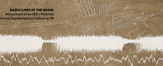
CINCINNATI – The potential for doctors to measure damaging “brain tsunamis” in injured patients without opening the skull has moved a step closer to reality, thanks to pioneering research at the University of Cincinnati Neuroscience Institute.
The research team, led by Jed Hartings, PhD, Research Associate Professor in the Department of Neurosurgery, has shown that spreading depolarizations — electrical disturbances that spread through an injured brain like tsunami waves — can be measured by the placement of electroencephalograph (EEG) electrodes on the scalp. The team demonstrated the effectiveness of the noninvasive technique by making simultaneous invasive recordings from electrodes that were placed directly on the surface of the brain in 18 patients who had undergone surgery following a traumatic brain injury.
“We can see clearly when spreading depolarizations occur with invasive recordings,” Dr. Hartings says. “And when we looked at this data side by side with the non-invasive EEG recordings, it just jumped out at us. When you look at the scalp EEG the right way, evidence of the spreading depolarization could be seen.”
The discovery has the potential to revolutionize bedside neuro-monitoring by enabling doctors to measure spreading depolarizations, which lead to worse outcomes, in patients who do not require surgery. At present, only about 10 to 15 percent of patients with TBI undergo surgery and are candidates for intracranial monitoring.
“This landmark observation broadens the spectrum of patients and conditions that can be monitored for the occurrence of spreading depolarizations,” says co-investigator Norberto Andaluz, MD, Mayfield Clinic neurosurgeon, Associate Professor of Neurosurgery and Director of the UC Neurotrauma Center at the UC Neuroscience Institute. Until now, spreading depolarizations could only be detected through the insertion of surgical leads into the patient’s brain. Thanks to this discovery, we will acquire a better understanding of the role that depolarizations play in virtually every neurological condition, thus opening new opportunities for therapeutic interventions.”
The research was published online in the journal Annals of Neurology earlier this month and highlighted online in the prestigious journal Nature Reviews. The research was funded by the U.S. Army’s Psychological Health and Traumatic Brain Injury (PH/TBI) Research Program and the Mayfield Education & Research Foundation.
The discovery was not made sooner because of historical conventions in how scalp EEG data is reviewed, Hartings says. Continuous EEG is typically studied in small 10-second segments, which allows detection of abnormal brain waves, such as epileptic spikes, which last fractions of a second. The changes associated with spreading depolarizations, on the other hand, develop over tens of minutes.
When the data is time-compressed so that hours of EEG recordings are presented on a single screen, the picture comes into focus. “The pattern jumps out at you,” Dr. Hartings says. “Suddenly, what you see developing over a course of 45 minutes is a depression of the amplitude of the brainwaves. It takes maybe 15 minutes for that to develop to its minimum, and then it will slowly recover again over another 15 to 20 minutes. So you can only see the depolarization when you are looking at long epochs of data.
“This is something that people have probably been recording for 50 years,” Dr. Hartings adds, “but they weren’t looking at the data in the right way to be able to recognize it.”
Dr. Hartings likened the phenomenon to the Nazca Lines, the famous geoglyphs in the desert of southern Peru. The Nazca lines suggest nothing up close; but when seen at a distance from surrounding foothills or an airplane, images of artistry emerge.
In the Annals of Neurology study, the researchers reported on spreading depolarizations that lasted from 10 minutes to several hours. More than 80 percent of the depolarizations could be detected with scalp EEG.
Bringing the “Nazca Lines of the brain” into focus from the patient’s bedside is a primary objective of Dr. Hartings’s research, which has played a leading role worldwide in the understanding of spreading depolarizations in acute neurologic injury. Dr. Hartings’s team characterized the phenomenon in TBI and laid the foundation for understanding its destructive nature in research published in Lancet Neurology and Brain in 2011.
“One of our main objectives going forward is to develop techniques for measuring spreading depolarizations non-invasively, using a more traditional EEG,” Dr. Hartings says.
Because the spreading depolarizations range from subtle to obvious, more research is needed. “If the activity is subtle, perhaps we can we use digital signal processing techniques to make the signal more obvious,” Dr. Hartings says. “Can we define data processing procedures that will clean up the signal and maybe even establish criteria that will set off an automatic alarm to alert bedside physicians?”
Translating and refining this knowledge into a broadly applicable methodology with a refined user-friendly bedside display, Hartings says, is perhaps five years away.
Additional co-investigators of the study included J. Adam Wilson, PhD, Jason Hinzman, PhD, Sebastian Pollandt, MD, Vince DiNapoli, MD, PhD, and David Ficker, MD. Dr. Ficker is a UC Health neurologist and Director of the EEG Laboratory and the Epilepsy Monitoring Unit at the UC Neuroscience Institute.
